
One of the most troublesome and poorly understood aspects of ME/CFS is the phenomenon known as post-exertional malaise (PEM). PEM refers to a sudden rapid energy loss that occurs after doing minor physical and/or mental tasks–there is literally no energy left, beyond what is needed for survival. This rapid decline in energy is difficult for family and friends to comprehend and they often have a hard time believing the condition is real. When normal people feel exhausted, they usually recover after a period of rest. This doesn’t happen to patients with ME/CFS, they cannot simply get out of bed and exercise–doing so only makes the their symptoms worse. This is because PEM is also marked by unpredictable flareups (e.g. cognitive impairments, increased muscle/joint pain, headache, etc.) can continue for hours or days, making it very difficult for patients to determine what events might have triggered their decline in function.
What it is like to live with PEM?
Here is a hypothetical case example. Jordan is a 35 year-old journalist suffering from ME/CFS for the past 2 years. One day she felt well enough to make plans to go out with her friends. When the time came, however, she suddenly felt completely drained with symptoms of lightheadedness, nausea, dizziness, extreme muscle weakness in her legs, intense joint pain, and hypersensitivity to noise, light, and temperature. She decided to cancel plans at the last minute to go lay down for several hours. The flareup became so intense that she could hardly make it to the bathroom. That night, she slept for 10 hours but felt unrested the next morning. She had planned to go grocery shopping, but was still down for the count. She tried to balance her checkbook but couldn’t concentrate to do the arithmetic. When her friend called to check in on her, she couldn’t follow the conversation and had difficulty finding the correct words to say. An email notification popped up on her phone, but she had to read it multiple times to understand the meaning. She was not sure what led up to this episode–she had avoided all the usual triggers. After 48 hours, her symptoms subsided enough that she felt she could resume her usual activities. However, she was now up against a deadline to write a news story she had been working on. The demands of her job meant she had to finish on time, only to crash afterward and become bedbound again. Because of this, she had to quit her job and do freelance writing. This allowed her to pace the work at her own schedule, but her output is very limited and she can barely cover living expenses.
What is the reason for PEM?
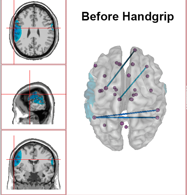
The underlying biological mechanisms for PEM are not fully understood. However, we have new preliminary findings using quantitative EEG with eLORETA which offer certain clues about PEM, suggesting it involves the autonomic nervous system and brain energy metabolism. Throughout the brain, there is a large network of regions called the central autonomic network that work together to coordinate activities of the body’s autonomic nervous system. We recorded the EEG of 7 patients with ME/CFS and 6 healthy controls immediately before and after doing a moderate handgrip exercise and recorded them again the next day to assess for changes that might occur after a 24-hour period. In the patient group, we saw a significant reduction in brain activity after exercise and it worsened after 24 hours. But in healthy control group, we saw the opposite pattern–there was a significant increase immediately after exercise, with further increase in activity after 24 hours. This makes sense from the standpoint of our autonomic nervous system, which is responsible for matching our physiology to ever-changing task demands. It’s job is to optimize blood circulation, body temperature, heart-rate and blood pressure, digestion, etc. from not only momentary exercise, but also in anticipation of exercise challenge.
The EEG is directly measuring neuronal activity occurring in milliseconds and each neuron has high metabolic energy consumption. The brain itself is a major energy challenge and there is an estimated 100 billion neurons that consume a disproportionate amount of energy–it accounts for up to 20 percent of the entire body’s energy budget, while being only 2% body weight. But neurons are highly sensitive to energy limitations. During this 300 millisecond time frame, the neurons in different regions of the brain are less active from insufficient energy production. Immune and metabolic processes also depend on one another and immune cells rely on energy (i.e., glucose) from the metabolic system to fight infections.
What are the implications for these new preliminary findings?
A much larger study is needed to confirm these results. However, these findings suggest that the central autonomic network may serve as an index or marker for PEM in some patients, which is the most debilitating feature of ME/CFS. Quantitative EEG (qEEG) could be used in the clinic for measuring subtle downregulation in brain function elicited by controlled exercise in order to capture PEM. These findings explain why patients are unable to exercise and why exercise can make symptoms even worse. People with ME/CFS often look fine on the outside while their autonomic system is failing to adapt cellular activities on the inside. PEM is largely based on patient self-report and 2-day cardiopulmonary exercise testing that many patients cannot actually do because they are too disabled.
qEEG can be used to provide objective findings of PEM showing that worsening symptoms are due to an underlying neurological condition that needs to be taken seriously. It is important that we examine all the biological reasons for the wide-ranging symptoms reported by patients with ME/CFS. When there are no major indications on routine tests, patients are typically told there is nothing wrong and suggestion is made that the problem is purely psychological. This is unfortunate because there may be follow-up tests for demonstrating autonomic dysfunction stemming from post-viral/immune responses in the brain. But we also need to develop new tests that with sufficient sensitivity for detecting the exercise-induced changes in PEM occurring both centrally and peripherally.
When there are no major indications on routine clinical tests, patients are typically told there is nothing wrong. People with ME/CFS have an invisible illness, where they may look fine on the outside while their body is failing to maintain steady-state on the inside. These objective findings help validate PEM, demonstrating that worsening symptoms are due to an underlying neurological condition that needs to be taken seriously. The NCRI is committed to the intensive study of the central autonomic network in relation to nearly all aspects of this debilitating disease.
Follow these links to read 2 separate interviews of these findings: ME Association’s guest blog and Phoenix Rising.
The full text article is available for free at: Central Autonomic Network Disturbance in Myalgic Encephalomyelitis/Chronic Fatigue Syndrome: A Pilot Study | 30 June 2021