
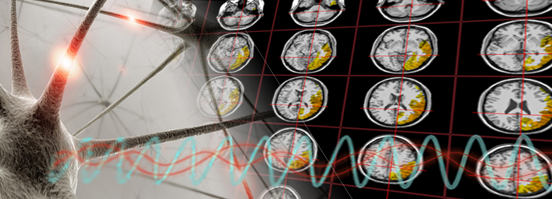
The NCRI promotes innovative brain research using the latest advancements in neuroimaging to inquire about mechanisms of the nature and extent of central nervous system involvement in neurological disorders. Here, we utilize eLORETA1, sLORETA or swLORETA, all of which derive their information from a quantitative electroencephalography (qEEG) record. If you are mathematically inclined, the method used to derive the originating sources of electrical activity within deeper brain structures is called the inverse solution. Using the latest state-of-the-art electrical neuroimaging methods, we can map areas of dysregulation, including large-scale networks, linking specific brain locations to patient symptoms and complaints. These integrative methods characterize the dysregulation found within brain systems which cause the disease process. In this manner, our applied research methods provide assessment, treatment monitoring, diagnosis, and observing the course of illness. Furthermore, this approach to neuroimaging is a fraction of the cost of fMRI and other modalities, so we can conduct research for a relatively small fraction of the cost involved in other neuroimaging modalities. Furthermore, electrical neuroimaging methods measure neuronal activity directly with high temporal resolution (timescale of milliseconds rather than seconds it takes using fMRI, PET, and SPECT).
Here at the NCRI, we focus on rare and poorly-understood neurological disorders as well as cognitive impairment resulting from illnesses, organ transplants, viral infections, critical illness, and other severe medical conditions. To develop better understandings and treatments for these disorders, we must look at diverse areas such as how movement is created, how the brain predicts the future, and why cognitive impairment does result from seemingly unrelated problems such as being in the ICU, having the flu or receiving a lung transplant. Neurological impairment can, and does, result from many types of insults. If there is a pathogen which travels to the brain, it does brain damage through the inflammation inherent in encephalitis.
We are also interested in how the brain compensates for neurological insults, including how the brain engages its compensatory mechanisms so as to continue working in the face of damage from disease or injury. Compensatory mechanisms attest to the brain’s incredible ability to re-work itself. Unfortunately, many patients who do have brain damage appear normal on neuropsychological tests, sometimes as a result of this compensation. Therefore, another research goal is to characterize brain damage separately from the brain’s normal compensatory abilities, allowing researchers to develop even more effective rehabilitation programs and helping those with brain damage.
References
- Pascual-Marqui, R. D., Lehmann, D., Koukkou, M., Kochi, K., Anderer, P., Saletu, B., … Kinoshita, T. (2011). Assessing interactions in the brain with exact low-resolution electromagnetic tomography. Philos Trans A Math Phys Eng Sci, 369(1952), 3768–3784. https://doi.org/10.1098/rsta.2011.0081
- Grech, R., Cassar, T., Muscat, J., Camilleri, K. P., Fabri, S. G., Zervakis, M., … Vanrumste, B. (2008). Review on solving the inverse problem in EEG source analysis. J Neuroeng Rehabil, 5, 25. https://doi.org/10.1186/1743-0003-5-25